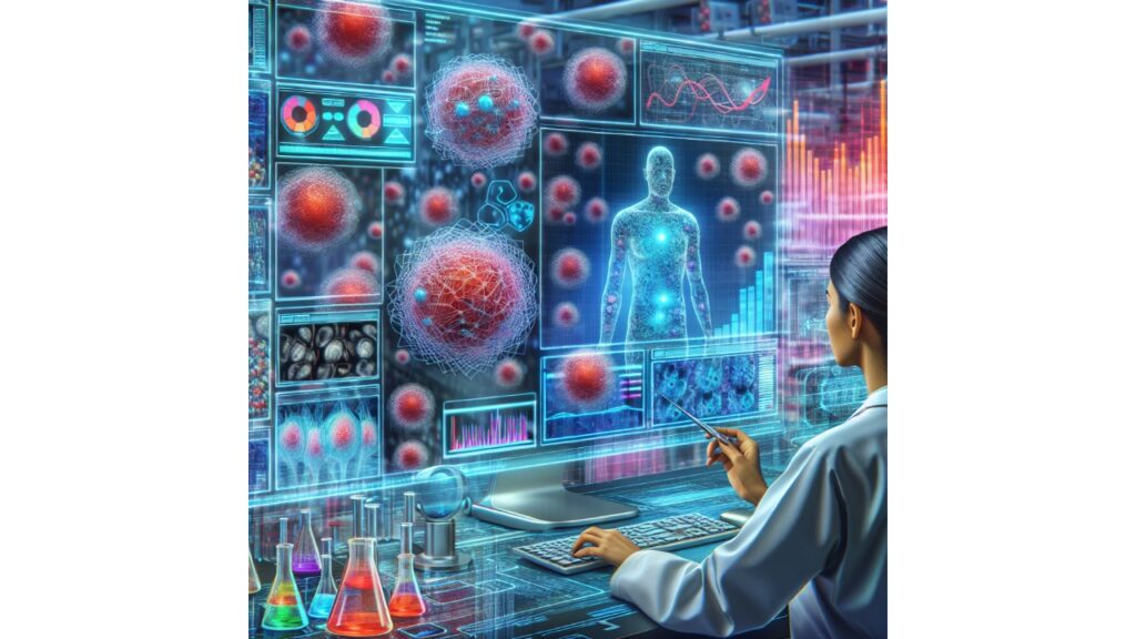
Stem cells function as the human body’s emergency tool kit. They have the unusual ability to differentiate into a variety of specialized cells, including immune cells and brain cells. They can divide and renew eternally, repairing and replenishing our system at will.
The Holy Grail of medicine is being able to cultivate stem cells in the lab and develop them into any kind of cell we require. For example, this capability would allow medical professionals to generate an infinite supply of fresh cells for repairing injured tissues and organs. But before we can find that Holy Grail, we must have a thorough grasp of how stem cells divide and differentiate into various cell types.
The Alfred E. Mann Department of Biomedical Engineering at USC has released new research that advances our understanding of these vital cells. Keyue Shen, an associate professor of biomedical engineering, and his colleagues have used machine learning to create a non-invasive device that provides a previously unobserved window into the process by which stem cells divide and regenerate into specialized cells.
Science Advances has published the work.
According to Shen, the activity of stem cells is still mostly unknown, and studying how they divide and alter requires the extraction and eventual destruction of stem cells in a lab.
The new study looks at hematopoietic stem cells, which develop into all the blood cells in our body, including immunological and red blood cells, and reside in our bone marrow. According to Shen, symmetric division of the stem cells is necessary for population expansion, while asymmetric division is necessary for self-renewal and the creation of a new cell type (like red or white blood cells).
When it comes to bone marrow transplants, ideally stem cells should to divide symmetrically to produce the greatest number of stem cells feasible, which can then utilize on various patients. However, according to Shen, blood stem cells in a clinic cannot actually be expanded outside of a body at this time. Many patients’ major problems will be resolved if they are able to produce a large quantity of hematopoietic stem cells for use in bone marrow transplants.
Shen’s team used fluorescent lifetime imaging microscopy, a real-time imaging technique, to examine the metabolic function of the stem cell—specifically, how it converts glucose into energy.
Autofluorescence, the term for the luminous material produced by stem cells, enables imaging to follow the cells’ metabolism. The function and transition of the cells are closely linked to this metabolism.
One of these molecules, NADH, for instance, is autofluorescent and exhibits various optical fluorescent features that are measurable when it forms a link with a metabolic enzyme. Thus, Shen explained, they can measure them non-invasively without harming the cells.
Shen and colleagues used a mouse model to extract fluorescent features from stem cell images and create a library of 205 metabolic optical biomarker features from individual stem cells, 56 of which were linked to hematopoietic stem cell differentiation.
The group was able to trace the behavior and differentiation of stem cells over time by creating a clustering map of them against non-stem cells using the machine learning technique. The method used a score to evaluate if the stem cells were dividing symmetrically or asymmetrically, and whether a daughter cell was likely a stem cell.
They are not eradicating the cells, which makes it really fascinating. All they are doing is taking pictures of the cell and then removing its features. That can provide us with a wealth of knowledge about them.
The group’s real-time method of studying stem cell metabolism will yield more fundamental insights that may help with drug development, innovative stem cell therapies, and regenerative medicine procedures involving the growth and replacement of human tissues, organs, and cells.
These days, there are additional uses, like cell therapy. Shen stated that researchers have been attempting to create several cell types, such as T cells, macrophages, and others, each with a unique set of applications in various illness scenarios.
For those who work with stem cells, this is an intriguing technological advancement because it makes it feasible to view a stem cell’s status in real time and follow each cell throughout time, something that is not now achievable.


