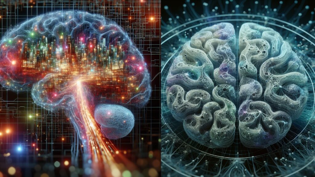
A tiny portion of the human brain has been meticulously mapped by researchers on an unprecedented scale, providing vivid details of individual brain cells, or neurons, and the complex networks they establish with other cells.
Approximately 57,000 neurons, 9 inches (230 millimeters) of blood vessels, and 150 million synapses—the sites where neurons connect—are visible on the ground-breaking brain map created by researchers from Google and Harvard.
The 10-year project’s co-leader, Harvard University professor of molecular and cellular biology Dr. Jeff Lichtman, said he was initially astounded by the intricate map. According to him, “I had never seen anything like this before,” he stated.
The 10-year project’s co-leader, Harvard University professor of molecular and cellular biology Dr. Jeff Lichtman, said he was initially astounded by the intricate map. According to him, “I had never seen anything like this before,” he stated.
With 86 billion neurons among its around 170 billion cells, the human brain is an incredibly complex organ. Using magnetic resonance imaging (MRI), researchers have previously peered into the brain at a millimeter scale. Furthermore, more recently, sophisticated microscopy methods have unveiled information at a far smaller scale, enhancing our comprehension of the inner workings of the brain.
Now, Lichtman and his colleagues have constructed a 3D map from a brain sample at the scale of a nanometer, or one millionth of a millimeter, utilizing these imaging techniques and an artificial intelligence (AI) technology called machine learning. This shows an image of the organ at the best resolution that science has yet produced.
Scientists can view the resulting cell atlas online as well; it was published in the journal Science on May 9.
A tiny portion of the brain, smaller than a rice grain, is depicted on this map. Its volume is around 1 cubic millimeter. A mature brain’s volume is a million times greater.
The woman, 45, had brain surgery to cure her epilepsy, and a sample of her brain was taken. The fragment was taken out of her brain by doctors from the cerebral cortex, which is the outermost area. The material was fixed in preservatives, and then heavy metal staining was applied to enhance the visibility of the cells. After that, they chopped the tissue into almost 5,000 slices, each of which was roughly 30 nanometers thick, and embedded it in resin.
That is roughly one-thousandth the thickness of a hair strand, according to Lichtman.The researchers used a high-speed electron microscope, which illuminates sample cells using multiple electron beams, to scan each slice. The microscope data was then forwarded to Google for additional AI processing.
Google’s scientists generated a three-dimensional (3D) representation of each object in all the microscopically captured photographs by using machine-learning algorithms to recognize the same thing in several images. After that, they electronically joined the renderings to create a three-dimensional reconstruction of the entire sample. One million gigabytes, or 1.4 petabytes, of data make up the final 3D map.
According to an email sent to Live Science by Viren Jain, a senior staff scientist at Google who co-led the study, the volume and complexity of the data gathered in this project demanded that Google be able to use cutting edge machine learning and artificial intelligence algorithms in order to reconstruct the 3D connectome.
There are several surprises in the scientists’ comprehensive map. For example, they discovered that certain axons, or the neurons’ outgoing lines, knotted themselves together to form whorls that Jain called both puzzling and beautiful. Additionally, the researchers discovered unusual neural connections in which a single axon could be connected to as many as 50 synapses.
These linkages may help to explain how quick reactions or significant memories are stored, though they are still learning more about how they work, Jain told.
If the whorls and extraordinarily strong connections are indicative of epilepsy in the tissue donor or if they would be observed in the brains of healthy individuals, Lichtman said it is unclear. To start answering the question, he said, the team is currently looking at brain tissue from a Parkinson’s patient.
Furthermore, he noted that because each person’s experiences have an impact on how their brain wires itself, it is doubtful that brain tissue samples from two different people will appear exactly the same.
A mouse’s brain is 500 times larger than a human brain sample, and that is the target of the team’s next task: mapping the complete brain. The hippocampus area, which is important for memory and learning, is where they are starting.
According to Lichtman, they have already started this challenging project.


