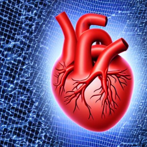
Cardiologists frequently utilize magnetic resonance imaging (MRI) and electrocardiograms (ECG) to map the structure of the heart and record its electrical activity, respectively. Physicians often do independent analyses of the two types of data to identify various heart-related facts.
In a new paper published in Nature Communications, researchers from the Eric and Wendy Schmidt Centre at the Broad Institute of MIT and Harvard have developed a machine learning method that can simultaneously learn patterns from ECGs and MRIs and predict characteristics of a patient’s heart based on those patterns. With further refinement, such a technology might one day aid medical professionals in identifying and diagnosing cardiac diseases more accurately from routine testing like ECGs.
The researchers also demonstrated that they could create MRI movies of the same heart using considerably more expensive imaging technology while still analysing ECG data, which are simple and inexpensive to obtain. Their approach might even be used to discover novel genetic markers for heart disease that current approaches that focus on particular data modalities might miss.
The researchers claimed that their technique offers a more comprehensive approach to understanding the heart and its diseases. According to Caroline Uhler, co-senior author of the study, member of the Broad core institute, co-director of the Schmidt Centre at Broad, and professor in the Department of Electrical Engineering and Computer Science as well as the Institute for Data, Systems, and Society at MIT, it is obvious that these two views, ECGs and MRIs, should be combined because they offer various perspectives on the condition of the heart.
The science of cardiology is fortunate to have a variety of diagnostic techniques, each of which offers a unique perspective on the physiology of the heart in both health and disease. Anthony Philippakis, a senior co-author on the paper and chief data officer at Broad as well as co-director of the Schmidt Centre, noted that one difficulty is that we lack systematic techniques for combining multiple modalities into a single, coherent picture. A initial step towards creating such a multi-modal characterization is represented by this work.
Model making
The researchers utilized a machine learning technology known as an autoencoder to create their model. Autoencoders automatically combine enormous amounts of data into a brief representation, or a more simplified version of the data. Following that, the group fed this representation into additional machine learning models to generate precise predictions.
In their research, the scientists first trained their autoencoder using heart MRIs and ECGs from UK Biobank users. Tens of thousands of ECGs were sent into the system along with identical MRI images. After that, the algorithm produced shared representations that integrated key information from both forms of data.
Adityanarayanan Radhakrishnan, a co-first author on the paper, an Eric and Wendy Schmidt Centre Fellow at the Broad and a graduate student at MIT in Uhler’s group, noted that once you have these representations, you can utilize them for a variety of applications. The other co-first author is Sam Friedman, a prominent machine learning scientist on the Broad’s Data Sciences Platform.
Predicting heart-related features is one of these uses. The researchers developed a model that could predict a variety of attributes, including aspects of the heart, such as the weight of the left ventricle, as well as other patient variables connected to heart function, such as age, and even heart illnesses. Furthermore, their model outperformed more conventional machine learning techniques as well as autoencoder algorithms that had only been trained on one imaging modality.
We demonstrated that if you use a variety of data sources, your forecast accuracy would increase, according to Uhler.
Because their model used representations that had been trained on a much larger dataset, Radhakrishnan explained that it was able to make predictions that were more accurate. Because autoencoders don’t need data that has been labelled by humans, the team was able to feed their autoencoder approximately 39,000 unlabeled pairs of ECGs and MRI images rather than just 5,000 labelled pairs.
By creating additional MRI movies, the researchers showed off another use for their autoencoder. The model produced the predicted MRI movie for the same person when an individual’s ECG recording was entered into it without a paired MRI recording.
The scientists believe that with further development, this technique could potentially allow doctors to understand more about a patient’s heart health from merely ECG recordings, which are regularly taken in doctors’ offices.
Additional gene searches
The scientists realized they could search for genetic variations linked to heart disease using their autoencoder representations. A genome-wide association study (GWAS), a common technique for identifying genetic variations for diseases, needs genetic information from people who have been diagnosed with the relevant condition.
The team was able to produce representations that indicated the general condition of a patient’s heart, however, because their autoencoder system doesn’t require labelled data. These illustrations and genetic information on the same patients from the UK Biobank were used by the researchers to build a model that searched for genetic variations that have a more general impact on the condition of the heart. The algorithm generated a list of variants that included many of the heart disease-related variants that are well-known as well as some new ones that may now be further examined.
According to Radhakrishnan, the autoencoder framework may have the greatest influence on genetic discovery, not just for heart disease but for all diseases, as more data and research are collected. Applying their autoencoder framework to the study of neurological diseases is something the research team is already engaged in.
This initiative, according to Uhler, is a good illustration of how machine learning researchers working alongside biologists and medical professionals can produce improvements in the interpretation of biomedical data. Making machine learning researchers interested in biomedical problems has the exciting potential to lead to completely new approaches to problems.


